
If you are looking at your profile and thinking that the area just above the tip looks heavy, rounded or “beak-like,” it is understandable to search for a quick fix. Revision planning becomes safer when the question changes from “How do I remove this bump?” to “What is creating the imbalance?” That distinction matters because removing more tissue from an already weakened nose can make a structural problem worse.
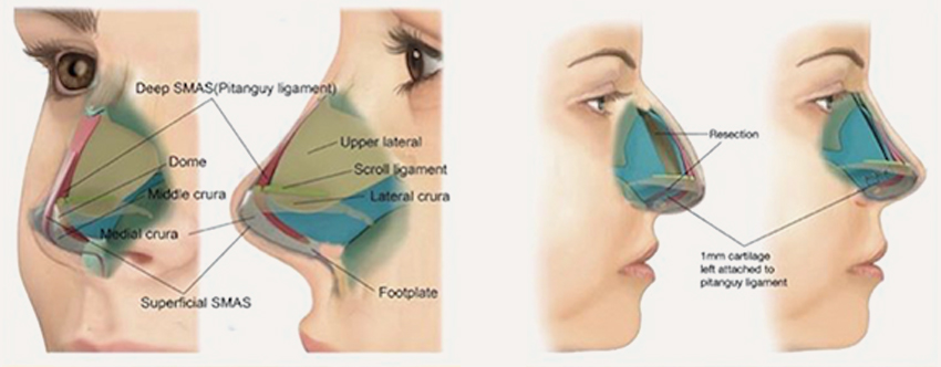
What Does “Pollybeak” Mean After Rhinoplasty?
Pollybeak deformity describes a convex or overly full supratip area that disrupts the normal relationship between the bridge, supratip and nasal tip. Patients may notice that the tip appears to point downward, the profile looks heavier than expected, or the upper tip never developed the definition they were hoping for. The visible shape is real, but it does not identify the underlying cause by itself.
Why Scar Tissue and Structural Pollybeak Need Different Plans
A scar-dominant supratip can look bulky even when the cartilage framework is reasonably positioned. In contrast, a structurally weak tip can make the supratip look prominent because the tip has lost projection or rotation. A low bony bridge can create a similar visual imbalance. Many revision patients have more than one factor at the same time.
This is why treatment cannot be chosen from one side-profile photograph. Timing, palpation, the quality of the skin-soft tissue envelope, breathing function, previous operative details and the remaining cartilage support all influence the plan. Early postoperative fullness may still be changing; a stable deformity a year later is a different clinical problem.
When Should Pollybeak Be Reassessed for Revision?
Revision rhinoplasty is usually considered only after the first operation has had enough time to heal and the shape has stabilized. In many cases that means waiting around a year before a new operation is planned. The reason is not a calendar rule for every patient: swelling and scar maturation can continue for months, and operating in actively healing tissue may make diagnosis and dissection more difficult.
- Seek earlier medical review if you have infection signs, skin compromise, severe obstruction, trauma or a sudden structural change.
- Do not assume a three- or six-month profile is the final result, especially in thick skin or revision tissue.
- A stable shape problem and a changing swelling pattern should not be managed as if they were the same condition.

How a Revision-Focused Assessment Separates the Cause
A useful assessment is systematic. The surgeon first maps the profile from the radix to the tip, then evaluates tip support, the cartilaginous and bony dorsum, skin thickness, scar behavior and nasal airflow. Front, three-quarter, profile and base views are compared rather than relying on the most flattering or most concerning angle.
- Review the date and type of every previous rhinoplasty or septorhinoplasty.
- Compare preoperative photographs with current standardized photographs.
- Identify whether the supratip fullness is soft, firm, mobile or fixed during in-person examination.
- Check tip projection and rotation rather than focusing only on the supratip.
- Assess internal and external nasal valve function and septal alignment when breathing symptoms are present.
- Determine whether remaining septal cartilage is available or whether reconstruction may require another graft source.
What Revision Surgery May Need to Correct—Without Prescribing a Technique Online
A true structural pollybeak may require the surgeon to restore a more balanced relationship between the tip and dorsum. Depending on anatomy, that can mean improving tip support, correcting dorsal imbalance, managing scar tissue carefully, or combining several maneuvers. The correct method is case-specific; the important principle is to avoid treating every pollybeak as “too much cartilage” or every supratip fullness as “just scar.”
What Real Case Evidence Should Show
For supratip fullness and pollybeak deformities, dynamic video assessment reveals whether the profile imbalance originates from excess soft tissue, cartilage over-projection, or loss of structural tip support.
Evaluating tip rotation, supratip break definition, and dorsal contour across dynamic angles provides critical insight into tissue elasticity and framework stability.
Before-and-after photographs are useful only when they are interpretable. For a pollybeak case, the most valuable comparison includes standardized profile and three-quarter views, the number of previous operations, the dominant diagnosis, whether breathing symptoms were present and the verified follow-up interval. A beautiful image without case context is marketing; a labeled case helps patients understand whether their problem is genuinely comparable.
What to Send for an Online Revision Assessment
A preliminary photo review can help determine whether your concern looks predominantly structural, soft-tissue related or mixed, but it cannot replace an examination. Send unfiltered front, both profile, three-quarter and base photographs; the date and location of your previous surgery; any operative report you have; a short description of breathing; and what specifically bothers you in the mirror or in photographs.
Questions Patients Commonly Ask Before a Revision Consultation
Some early soft-tissue fullness may continue to change as swelling and scar mature. Certain nonsurgical measures may be considered by a treating physician in selected cases. A stable structural deformity, however, requires a different discussion. The key is determining which situation you have.
No. Research and revision practice describe several mechanisms, including excessive supratip scar, inadequate tip support and imbalance between the bony dorsum and tip. More than one mechanism may be present.
Not necessarily. Graft choice depends on the amount and quality of cartilage remaining, how much support must be rebuilt and whether the nose has had multiple previous operations. That decision should follow examination and surgical planning, not a keyword or photograph alone.
The Next Step Is a Diagnosis, Not a Promise
Revision rhinoplasty is most useful when the problem is clearly defined and the patient understands what can and cannot be changed. If your main concern is a heavy supratip or downward-looking profile after a previous rhinoplasty, start by documenting the problem in standardized photographs and sharing your prior surgical history. The goal of the first review is to decide what needs to be examined—not to guarantee an outcome from a screen.
Why the Same Pollybeak Shape Can Mean Different Things
A pollybeak is a visual description, not a complete diagnosis. Two patients can have a very similar profile and still need completely different discussions. In one nose, the framework may be strong and the visible fullness may be driven mainly by thickened scar tissue. In another, the tip may have lost projection so the supratip appears relatively dominant. A third patient may have a bridge that was reduced more than intended, making the supratip look fuller even when there is no true excess in that area. Many complex cases combine more than one of these mechanisms.
This distinction matters because revision surgery is not simply a matter of removing whatever looks prominent. A surgeon has to understand the relationship between the bridge, supratip, tip support, skin-soft tissue envelope and airway. The safer question is therefore not “How can the bump be shaved down?” but “Why does this profile look unbalanced, and what must be preserved or rebuilt to improve it?”
How Skin Thickness and Scar Behavior Change the Visual Result
Skin thickness can strongly influence how the supratip behaves after rhinoplasty. Thicker skin may hide fine structural definition and can remain swollen or firm for longer. Thin skin, on the other hand, can reveal small framework irregularities more clearly. Neither type is automatically “better” or “worse”; each changes how the underlying structure is seen and how postoperative changes are interpreted.
Scar tissue also changes over time. Early firmness or fullness can soften as healing progresses, while a mature, stable contour that has persisted after the healing period raises a different set of questions. For this reason, serial photographs taken under similar lighting and from the same angles are more informative than comparing isolated selfies taken with different lenses, distances or head positions.
What a Realistic Revision Goal Looks Like
A realistic revision goal is not to create an artificial “perfect” profile line. It is to restore a more coherent relationship between the dorsum and tip while respecting skin quality, remaining cartilage, scar planes and breathing function. Some patients need a small contour correction; others need structural support to be rebuilt before the profile can look balanced. The amount of possible change is therefore individual.
A strong consultation should explain the dominant problem, the secondary contributing factors, the limitations created by the first operation and the expected healing process after revision. It should also make clear what cannot be promised. In a previously operated nose, stable improvement is more important than aggressive reduction.
Questions Worth Asking During a Revision Consultation
- Is the supratip fullness mainly scar-related, structural, or mixed?
- Has tip support been reduced, and does projection need to be restored rather than reduced further?
- Is the bridge height contributing to the pollybeak appearance?
- Are there breathing or nasal-valve issues that should be assessed at the same time?
- What evidence from my old photographs or operative records helps clarify what changed?
- What are the realistic limits of correction in my skin and scar pattern?
Additional Patient Questions
Photographs can suggest a pattern, but they cannot reliably show tissue firmness, internal anatomy, scar planes or dynamic support. They are useful for preliminary screening and comparison, while the final distinction requires examination.
Yes. As generalized postoperative swelling settles, the relationship between the supratip and tip can become easier to see. In some cases the profile appears more balanced with time; in others a stable structural imbalance becomes clearer. That is one reason timing and serial documentation matter.
No. Revision complexity depends on the cause, the amount of previous structural change, scar quality and the correction required. Some cases are limited; others require reconstruction. The word “revision” does not define the size of the operation by itself.
Pollybeak Revision Consultation
Connect directly with Dr. Emre İlhan Clinic coordinators on WhatsApp (+90 541 379 64 94) to evaluate your photos and case history.
Request a Personalized Revision Assessment
A consultation becomes precise when our surgical team receives clinical details regarding your surgical history, breathing symptoms, and primary aesthetic goals.
Request a Revision Assessment
Connect directly with Dr. Emre Ilhan’s surgical coordination team via WhatsApp for confidential pre-evaluation and image review.